Tonsil and Adenoid Anatomy Biology Diagrams Pharyngeal tonsil (medial view) The adenoid is a pyramidal shaped structure composed of lymphoid tissue. The apex of this pyramid is extended towards the to nasal septum, and the base sits at the posterior most wall of the nasopharynx. The adenoidal surface is invaginated by a number of folds with some crypts.There is a midline pharyngeal bursa (bursa of Luschka) which extends posteriorly and
The pharyngeal tonsil, also known as the adenoids, is the most superior component of the pharyngeal lymphoid ring and lies in the superior part (vault) of the nasopharynx.It is attached to the periosteum of the sphenoid bone by connective tissue.The pharyngeal tonsil is covered with ciliated pseudostratified columnar (i.e. respiratory) epithelium. The covering capsule is thinner compared to
Anatomy and physiology of the palatine tonsils, adenoids, and lingual ... Biology Diagrams
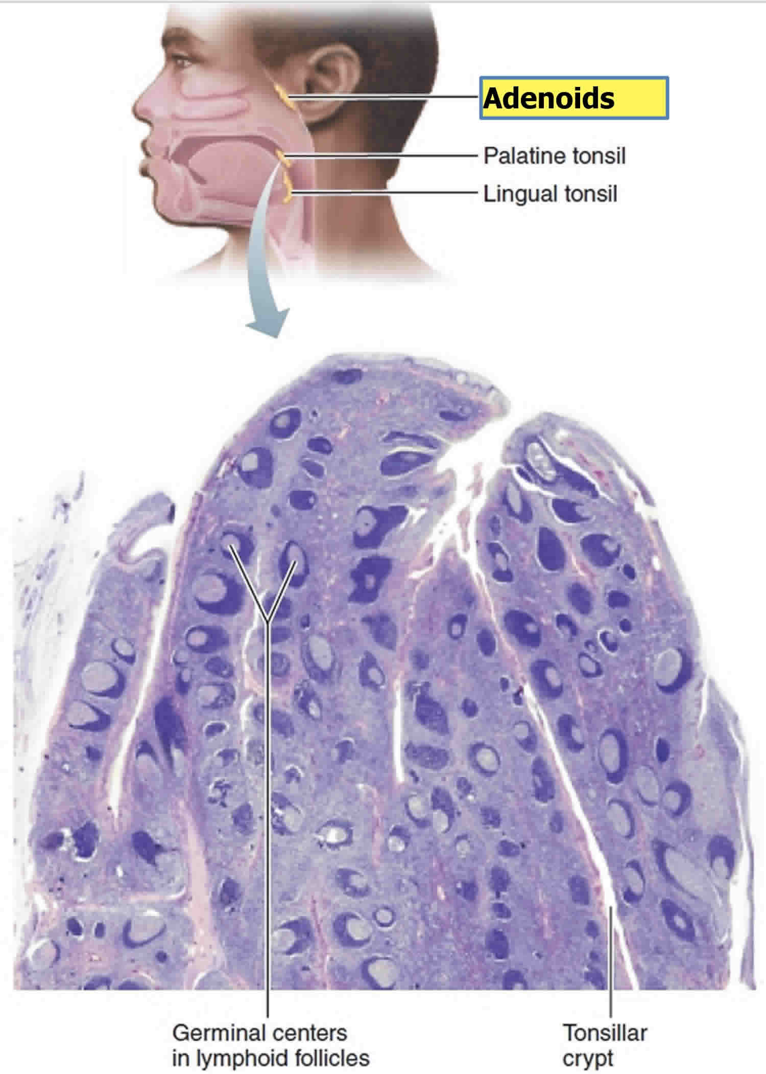
Keywords: Anatomy of tonsils, Adenoids, Waldeyer's ring. The palatine tonsils, adenoids, tubal tonsils, and lingual tonsils are lymphoepithelial tissues that make up the components of Waldeyer's ring, named after the German anatomist Heinrich Wilhelm Gottfried von Waldeyer-Hartz. These entities are together a part of the mucosal immune system. The adenoids exist as a rectangular mass of lymphatic tissue in the nasopharynx. Meyer first described this mucosa-associated lymphoid tissue in 1868. The adenoids are midline structures situated on the roof and posterior wall of the nasopharynx. They form part of the Waldeyer ring, whose components include the adenoids, the palatine tonsils, and the lingual tonsils. They are present from the
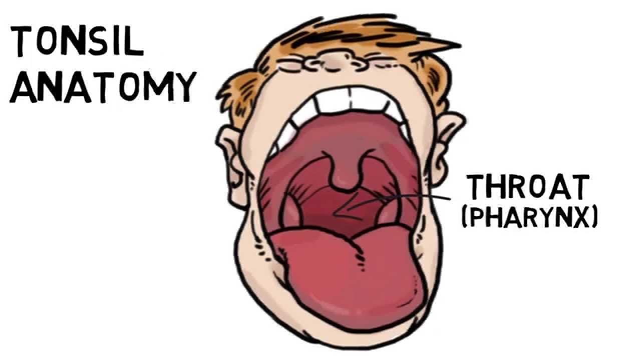
Tonsils are lymphoid tissue aggregates situated near the entrance of the digestive and respiratory tracts and play a key role in our immune system. They act as a front-line defense forming the initial immunological response to inhaled or ingested pathogens. The lymphatic tissues located in the oropharynx are composed of a circumferential tonsillar ring, known as the Waldeyer's ring which

Anatomy and Physiology of Tonsils Biology Diagrams
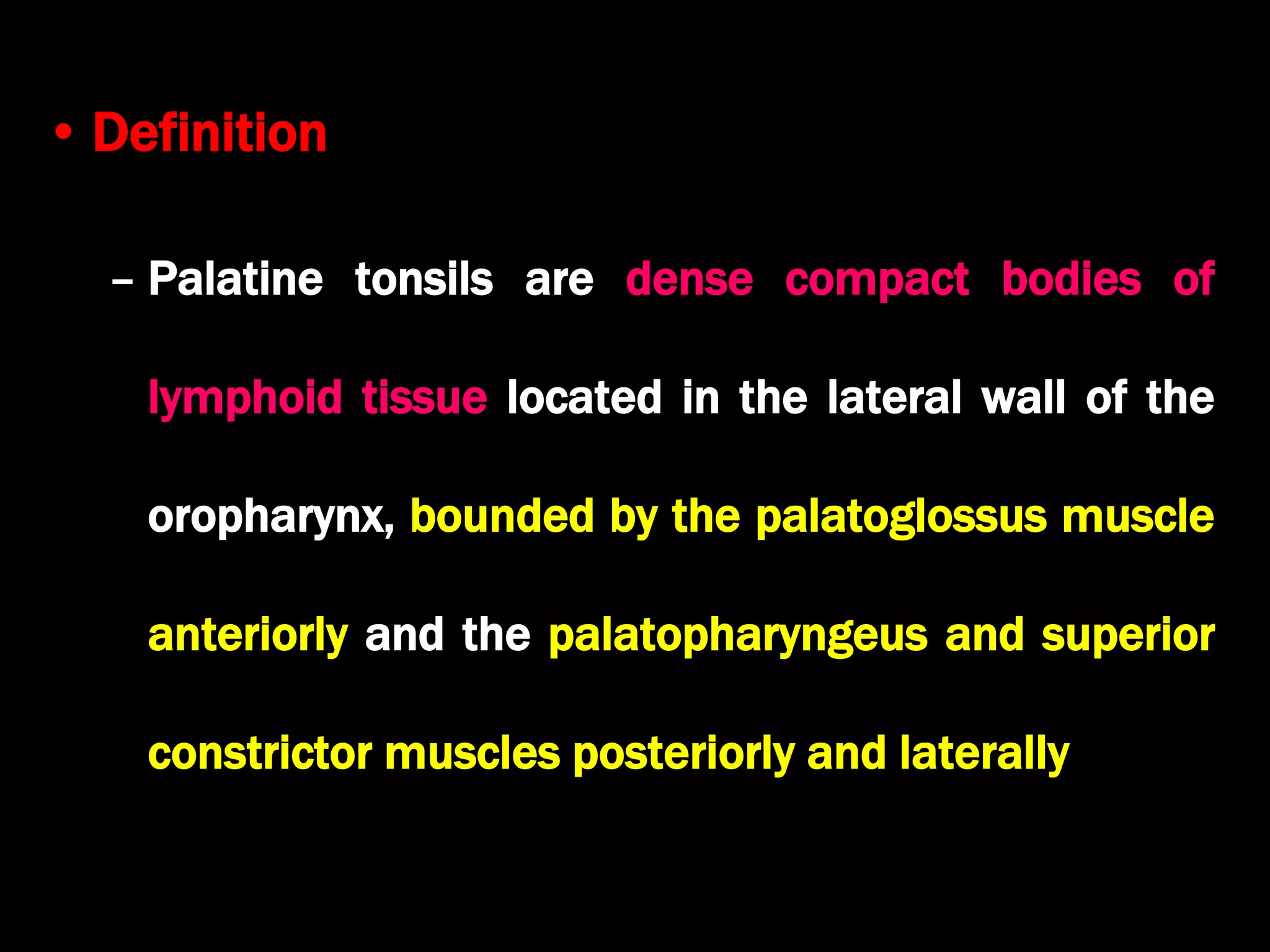
Objective: This review aims to discuss the basic anatomy and physiology of the palatine and pharyngeal tonsils, with reference to how this foundational understanding may affect patient management and surgical procedures in these regions of the upper airway. Methods: A literature search was performed using PubMed and Google Scholar using the MeSH terms tonsils, adenoids, anatomy, physiology The palatine tonsils are dense compact bodies of lymphoid tissue that are located in the lateral wall of the oropharynx, bounded by the palatoglossus muscle anteriorly and the palatopharyngeus and superior constrictor muscles posteriorly and laterally. The adenoid is a median mass of mucosa-associated lymphoid tissue.
