ligament attachments anterior Printable Worksheet Biology Diagrams This ligament has two attachment points; a proximal attachment on the medial condyle of the tibia, and a distal attachment on the medial shaft of the tibia. Anteriorly, the superficial part blends with the medial patellar retinaculum and the medial patellofemoral ligament, which courses from the medial femoral condyle to attach onto the medial
It is caused by axial traction or a sudden pull of the extended pronated arm. The radial head moves out of the weak annular ligament and capitellum, resulting in slipping over and subluxation of the radial head into the supinator muscle and annular ligament. Assessment [edit | edit source] Typical history might include a sharp jerk to the arm. Unlike other ligaments or tendons, the anterior cruciate ligament normally has a heterogeneous appearance and the anteromedial and posterolateral bundles are defined by surrounding high-intensity structures 1.. The ACL Blumensaat line angle is normally ≤15º. It is calculated by drawing a line parallel to the roof of the intercondylar notch of the femur (Blumensaat line) and one parallel to

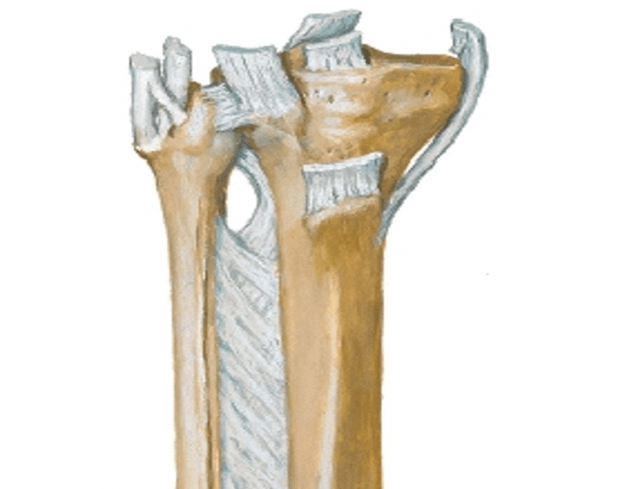
Annular Ligament Biology Diagrams
Inguinal ligament (Ligamentum inguinale) The inguinal ligament (also ligamentum inguinale, arcus inguinalis or Pouparts's ligament) is a band of connective tissue that extends from the anterior superior iliac spine of the ilium to the pubic tubercle on the pubic bone.. It is formed by the free inferior border of the aponeurosis of the external oblique muscle which attaches to these two points.

The anterior cruciate ligament (ACL) is a band of dense connective tissue which courses from the femur to the tibia. It consists of type I Both the medial and lateral intercondylar tubercles of the tibia serve as the attachment points for the two anteromedial and posterolateral bundles of the ACL.. For more detail on the anatomy of the

Anterior Cruciate Ligament (ACL) Biology Diagrams
The inguinal ligament or Poupart's ligament formed from the aponeurosis of the lower border of external obliquis muscle. It has 2 surfaces concave and convex, the convex surface toward the thigh attached to the deep fascia that pulling the ligament downward. It is a site for attachment of internal obliquis muscle at the lateral 2/3 and transversus abdominis at the lateral 1/3.
